Which Cones Are Activated for the Brain to Perceive Blue
![]()
| |
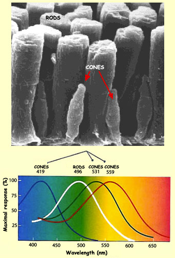
| | The two kinds of photoreceptors in the retinas of vertebrates—the rods and cones—differ in many ways, both anatomically and functionally. The main difference is the opposite roles that they play in vision. The rods provide what is called scotopic vision: they are very sensitive to low levels of light but cannot distinguish colours. The cones provide photopic vision: they require bright light but let us see the world around us in colour, and more sharply. In both cases, however, the neural response is the same—the hyperpolarization of the photoreceptor cells—and is initiated by the same phenomenon: the absorption of light energy by photopigments embedded in the discs of the photoreceptors' outer segments. In the rods, the photosensitive pigment is rhodopsin, which has its peak sensitivity at around 500 nanometres (nm) in the visible-light band of the electromagnetic spectrum.
But the attachment of color names to cones can be misleading because the cones are not maximally sensitive exactly in the red, green and blue parts of the spectrum. As you can see on the picture above, the blue cones are most sensitive in the violet, and the red cones in the yellow-green part of the spectrum. Consequently, it is more accurate to refer to the three types of cones containing mostly blue-, green-, and red-sensitive pigment as S-cones, M-cones, and L-cones, respectively, where S, M, and L stand for short, medium, and long wavelength. An object whose colour falls anywhere in the visible spectrum will therefore excite all three types of cones to varying extents. For example, a green object will stimulate green cones for the most part, but also red cones to a lesser extent, and blue cones to a still lesser one. Our perception of colours thus depends on this superimposition of the various absorption spectra of the three types of cones, and subsequently, of course, on the complex neuronal interactions between the retina and the rest of the brain. Colour blindness, or daltonism, is a vision defect characterized by the inability to differentiate certain colours or hues. Its name comes from that of the English physicist John Dalton (1766-1844), who suffered from this condition himself. About 8% of all men are colour blind to varying degrees, and slightly less than 1% of all women. The reason for this difference is that the main form of colour blindness is hereditary, and the genetic mutations that cause it occur on the X chromosome. Since the mutated gene is recessive, women, who have two X chromosomes, can carry the gene without being colour blind, if the other X chromosome is unaffected. But men have only one X chromosome, so if the mutated gene is present on it, they are automatically colour blind. Cases of total colour blindness, known as achromatopsy, in which someone sees the world only in shades of grey, are very rare. Usually, people who are colour blind have trouble in telling red from green, or, much more rarely, blue from yellow. Classic red/green colour blindness is the result of a lack of red cones in the retina. Forms of colour blindness are usually classified according to the type of cone affected. Thus there are three kinds of colour blindness, corresponding to the three kinds of cones. Blindness to green, due to deficiency of the green pigment, is called deuteranopia, and is the most common form. | |
| |
Dark adaptation is a two-step process in which the eyes make the transition from photopic (cone-based) to scotopic (rod-based) vision. Once you have spent a certain amount of time in a well-lit room, your eyes' light-sensitivity threshold is very high. If you then move into a darker room, this threshold falls rapidly for the first 5 or 6 minutes, then seems as if it were going to stabilize asymptotically. But around the 7th minute, the threshold starts to fall even more. About half an hour later, it reaches a second asymptotic level, much lower than the first. This minimum level is the threshold for scotopic vision, whereas the initial level represented the threshold for photopic vision. | |
The Vitamin A that our bodies produce from the beta carotene in many of the foods we eat (including, most famously, carrots) is needed to synthesize the retinene bound to the centre of the rhodopsin molecule. Indeed, a severe Vitamin A deficiency impairs night vision, because of the smaller amounts of retinene being produced. During the daytime, however, there is generally enough light to allow relatively normal vision despite low levels of visual pigments. | |
![]()
![]()
Source: https://thebrain.mcgill.ca/flash/a/a_02/a_02_m/a_02_m_vis/a_02_m_vis.html


0 Response to "Which Cones Are Activated for the Brain to Perceive Blue"
Postar um comentário